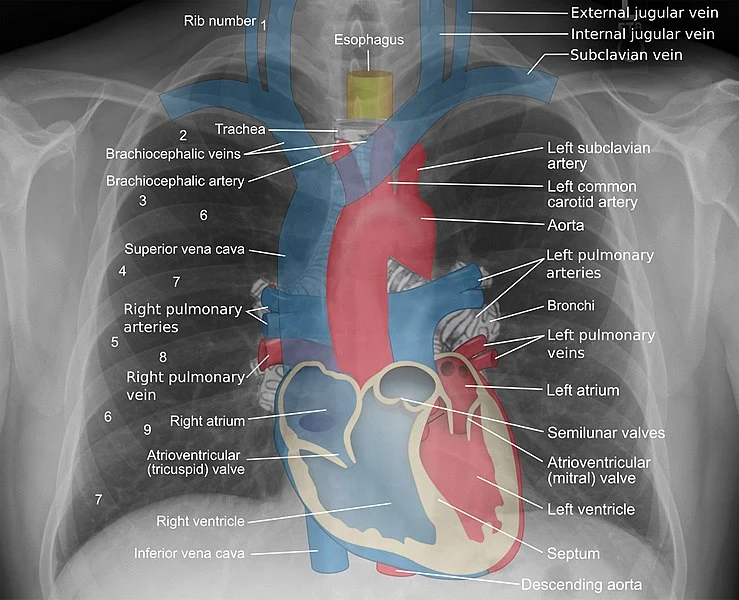
Anatomy Of Chest : Anatomy For Radiology Chest Glass Box. Make learning anatomy enjoyable, clear and fun. Browse 2,531 female chest anatomy stock photos and images available, or start a new search to explore more stock photos and images. Of the two chest muscles, the pectoralis major (a.k.a. Plus, how to target each to make them bigger and stronger. An overview of the anatomy visible in a transverse computed axial tomographical image of the thorax (and part of the abdomen) performed with intravenous cont.
This atlas is a comprehensive and affordable learning tool for medical students and residents and especially for radiologists and pneumologists. The chest anatomy includes the pectoralis major, pectoralis minor and the serratus anterior. The chest is the area of origin for many of the body's systems as it houses organs such as the heart, esophagus, trachea, lungs, and thoracic diaphragm. A good radiologist knows the anatomy because knowing where structures normally live and recognizing the location of an abnormality helps to make or narrow the differential diagnosis. Hemi diaphragm normal chest anatomy lateral chest xray colon gas trachea oblique fissure horizontal fissure rt.
Anatomy For Radiology Chest Glass Box from glassboxmedicine.files.wordpress.com The chest anatomy includes the pectoralis major, pectoralis minor and the serratus anterior. It provides access to ct images in the axial plane, allowing the user to learn and review the lung anatomy interactively. A typical heart is approximately the size of your fist: This chapter is an abbreviated review of thoracic anatomy as seen on chest radiographs and computed tomography (ct) of the chest. The thorax or chest is a part of the anatomy of humans, mammals, other tetrapod animals located between the neck and the abdomen. (1) the pectoralis major, and (2) the pectoralis minor. To carry out the unique functions performed by the chest wall, the anatomic structures are formed precisely for maximal efficiency. Explore with online courses on anatomy app.
Whatever you need, whatever you want, whatever you desire, we provide.
The chest is the area of origin for many of the body's systems as it houses organs such as the heart, esophagus, trachea, lungs, and thoracic diaphragm. Learn about each of these muscles, their locations, functional anatomy and exercises for them. The chest or thorax is the region between the neck and diaphragm that encloses organs, such as the heart, lungs, esophagus, trachea, and thoracic diaphragm. The chest anatomy includes the pectoralis major, pectoralis minor and the serratus anterior. The chest wall is a complex system that provides rigid protection to the vital organs such as the heart, lungs, and liver; A line is drawn from anterior surface of the body of 6th thoracic vertebrae passing through the apex of the heart up to anterior lower most part of diaphragm. A good radiologist knows the anatomy because knowing where structures normally live and recognizing the location of an abnormality helps to make or narrow the differential diagnosis. This atlas is a comprehensive and affordable learning tool for medical students and residents and especially for radiologists and pneumologists. Your sternum is a bone that's located in the middle of your chest. Chest a man's chest — like the rest of his body — is covered with skin that has two layers. As with all parts of the body, the anatomy and physiology of the chest wall are intimately intertwined. To carry out the unique functions performed by the chest wall, the anatomic structures are formed precisely for maximal efficiency. Download my two educational text books for free using this link:
Understanding chest wall anatomy is paramount to any surgical procedure regarding the chest and is vital to any reco. And flexibility to aid in the functional process of respiration. The chest or thorax is the region between the neck and diaphragm that encloses organs, such as the heart, lungs, esophagus, trachea, and thoracic diaphragm. Make learning anatomy enjoyable, clear and fun. An overview of the anatomy visible in a transverse computed axial tomographical image of the thorax (and part of the abdomen) performed with intravenous cont.
Shoulder And Chest Anatomy Artwork Stock Image C020 0121 Science Photo Library from media.sciencephoto.com The major muscle in the chest is the pectoralis major. Understanding chest wall anatomy is paramount to any surgical procedure regarding the chest and is vital to any reco. The chest or thorax is the region between the neck and diaphragm that encloses organs, such as the heart, lungs, esophagus, trachea, and thoracic diaphragm. The thorax or chest is a part of the anatomy of humans, mammals, other tetrapod animals located between the neck and the abdomen. It is enclosed by the ribs, the vertebral column, and the sternum, or breastbone, and is separated from the abdominal cavity (the body's largest hollow space) by a muscular and membranous partition, the diaphragm. The chest or thorax region of the upper body has a number of important organs that reside within it that may present with chest pain if they become compromised in. Download my two educational text books for free using this link: To carry out the unique functions performed by the chest wall, the anatomic structures are formed precisely for maximal efficiency.
It's also sometimes referred to as the breastbone.
As with all parts of the body, the anatomy and physiology of the chest wall are intimately intertwined. The epidermis is the outermost layer that provides a protective, waterproof seal over the body. The chest is made up primarily of two muscles: The muscles of the chest develop from the somites found in the mesoderm. And flexibility to aid in the functional process of respiration. This chapter is an abbreviated review of thoracic anatomy as seen on chest radiographs and computed tomography (ct) of the chest. Having to do with the chest. (1) the pectoralis major, and (2) the pectoralis minor. Get the full built by science program: The chest anatomy includes the pectoralis major, pectoralis minor and the serratus anterior. Stability to arm and shoulder movement; Learn about each of these muscles, their locations, functional anatomy and exercises for them. A good radiologist knows the anatomy because knowing where structures normally live and recognizing the location of an abnormality helps to make or narrow the differential diagnosis.
The chest is the area of origin for many of the body's systems as it houses organs such as the heart, esophagus, trachea, lungs, and thoracic diaphragm. Radiology basics of chest ct anatomy with annotated coronal images and scrollable axial images to help medical students and junior doctors learning anatomy. Your sternum protects the organs of your torso from injury and also serves as a. The major muscle in the chest is the pectoralis major. The chest wall is a complex system that provides rigid protection to the vital organs such as the heart, lungs, and liver;
Still S Disease And Muscle Movement Physics Forums from www.physicsforums.com (1) the pectoralis major, and (2) the pectoralis minor. The chest or thorax region of the upper body has a number of important organs that reside within it that may present with chest pain if they become compromised in. Summary:for adequate treatment of patients with breast cancer, mastologists should have a complete understanding of the anatomy of the thoracic wall, axilla and breast. As with all parts of the body, the anatomy and physiology of the chest wall are intimately intertwined. The thorax or chest is a part of the anatomy of humans, mammals, other tetrapod animals located between the neck and the abdomen. Anatomy of right side chest pain. The major muscle in the chest is the pectoralis major. This article focuses on the unique structural characteristics in …
This chapter is an abbreviated review of thoracic anatomy as seen on chest radiographs and computed tomography (ct) of the chest.
The shape of the chest is often regarded as potential insight into a disease process, as in the case of barrel chest and respiratory dysfunction. An overview of the anatomy visible in a transverse computed axial tomographical image of the thorax (and part of the abdomen) performed with intravenous cont. Learn about each of these muscles, their locations, functional anatomy and exercises for them. Anatomy of the thorax, heart, abdomen and pelvis recommended text gray's anatomy for students. About the 6th week, the somites differentiate into the sclerotomes and the dermatomyotomes. Whatever you need, whatever you want, whatever you desire, we provide. Stability to arm and shoulder movement; Here's how science can help you grow! The pec major) is the one that commands the most real estate. It's also sometimes referred to as the breastbone. Computed tomography (ct) of the chest can detect pathology that may not show up on a conventional chest radiograph(1). (1) the pectoralis major, and (2) the pectoralis minor. Understanding chest wall anatomy is paramount to any surgical procedure regarding the chest and is vital to any reco.
Share :
Post a Comment
for "Anatomy Of Chest : Anatomy For Radiology Chest Glass Box"


Post a Comment for "Anatomy Of Chest : Anatomy For Radiology Chest Glass Box"